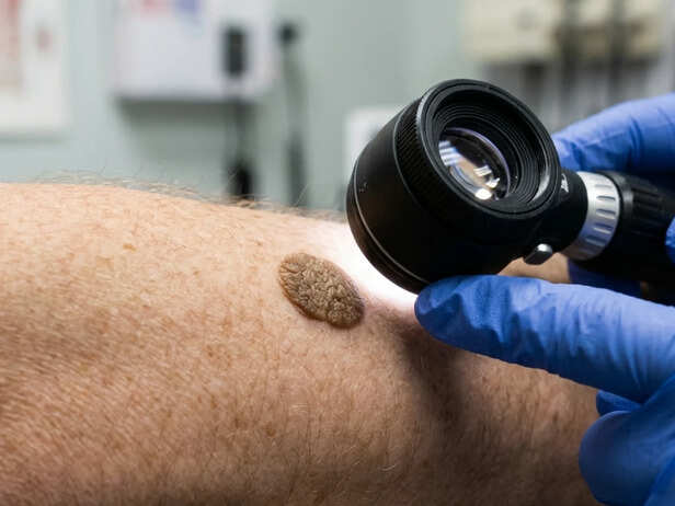
A stuck-on, darkened patch of skin that appears without warning is one of the fastest routes to health anxiety. Seborrhoeic keratoses are benign epidermal growths, not cancerous, not contagious, not caused by sun damage in the way melanoma is, yet they account for a significant share of dermatology referrals driven by patient concern. Understanding what they are, and what they are not, matters because misidentifying them leads to either unnecessary panic or, in rarer cases, missing something that does need attention.
What Is Actually Happening in the Skin
![Seborrhoeic keratosis treatment]()
Seborrhoeic keratosis treatment
How to identify seborrhoeic keratosis
Seborrhoeic keratoses originate in the keratinocytes, the cells that make up the outermost layer of the skin, the epidermis. For reasons that remain incompletely understood, clusters of these cells begin proliferating in a disorganised way, stacking up rather than shedding normally. The result is a thickened, raised lesion that looks as though it has been pressed onto the surface rather than grown from within it. That stuck-on appearance is one of the most reliable visual cues clinicians use.
They are not related to the sebaceous glands despite the name, and they are not warts, which are caused by human papillomavirus. Seborrhoeic keratoses have no viral origin. They tend to appear from the mid-thirties onward, become more numerous with age, and run in families, which points to a genetic component. Friction-prone areas, the chest, back, face, and scalp, are common sites, though they can appear almost anywhere except the palms and soles.
How Dermatologists Distinguish Them From Concerning Lesions
A clinical diagnosis of seborrhoeic keratosis requires examination by a dermatologist. This is not a condition to self-diagnose from a photograph or a symptom checklist. Incorrectly self-treating certain pigmented lesions, assuming they are benign when they are not, can delay the detection of melanoma or other skin cancers. The two can look similar to an untrained eye, particularly when a seborrhoeic keratosis is darkly pigmented or has an irregular border.
Dermatologists use dermoscopy, a handheld device that magnifies the skin surface under polarised light, to examine the internal structure of a lesion. Seborrhoeic keratoses have characteristic features under dermoscopy: comedo-like openings, milia cysts, and a cerebriform or gyrate surface pattern. Melanoma has a different set of dermoscopic signatures. When there is genuine uncertainty, a biopsy provides a definitive answer. A dermatologist who cannot confidently exclude a malignant lesion on clinical and dermoscopic grounds will not simply reassure and discharge, they will take a sample.
The practical implication: any pigmented or raised lesion that is new, changing in size or colour, bleeding, or itching persistently needs professional assessment. The fact that seborrhoeic keratoses are common does not make every dark patch one.
Who Gets Them and What Triggers Proliferation
Age is the strongest predictor. Most people will develop at least a few by their fifties; some develop dozens. A sudden eruption of multiple lesions, known as the sign of Leser-Trélat, can occasionally signal an underlying internal malignancy, though this is rare and the association remains debated in the literature. A dermatologist assessing a patient with a sudden, large crop of new lesions may consider whether further investigation is warranted, depending on the clinical picture.
Hormonal changes, pregnancy, and prolonged friction from clothing or skin folds are associated with their appearance, but none of these causes them in a mechanistic sense that is fully established. Ultraviolet exposure does not cause seborrhoeic keratoses the way it causes actinic keratoses, a genuinely pre-cancerous lesion that is a separate entity and requires different management.
Removal: When It Makes Sense and When It Does Not
Because seborrhoeic keratoses are benign, removal is almost never medically necessary. The exception is when a lesion cannot be distinguished from a malignant growth even after dermoscopy, in which case excision serves a diagnostic purpose as much as a therapeutic one.
Cosmetic removal is a separate conversation. Options include cryotherapy, in which liquid nitrogen freezes the lesion; curettage, in which it is scraped away after local anaesthetic; and certain laser or electrosurgical techniques. Each approach has a realistic range of outcomes: most lesions respond well to a single session, but pigmentation changes, either hypopigmentation or hyperpigmentation, are a recognised side effect, particularly in darker skin tones. People with Fitzpatrick skin types IV through VI should discuss this risk explicitly with their dermatologist before proceeding.
Home removal attempts using over-the-counter acids, hydrogen peroxide products, or physical scraping are not appropriate. They carry a real risk of scarring, infection, and, critically, of treating a lesion that was not actually benign without anyone having looked at it properly.
The decision to remove a seborrhoeic keratosis belongs to the individual, made with accurate information from a qualified dermatologist who has examined the lesion. No online tool, photograph comparison, or symptom list substitutes for that assessment. What this piece can offer is the framework for that conversation, not a conclusion about what any particular growth is.
Discover expert advice and the latest updates in
Skin Care,
Hair Care,
Wellness,
Dermatology, and more at
Stay Young guide to lasting beauty and holistic health!